Since 1994, Virginia Eye Center, P.C. has been delivering state-of-the-art eye care to Loudoun County and surrounding areas. In an era where old fashioned "bedside manner" is hard to find, our true passion is for our patients. We strive to embody our EPICC core values- Excellence, Positivity, Integrity, Compassion and Collaboration. In addition, our board-certified doctors have successfully completed thousands of surgical procedures and are well known for their surgical excellence. We use the latest equipment, technology and minimally invasive techniques to provide exceptional medical and surgical care including treatment of cataracts, glaucoma, dry eye, general eye diseases and contact lens fittings. Our goal is to deliver an EPICC experience to you!
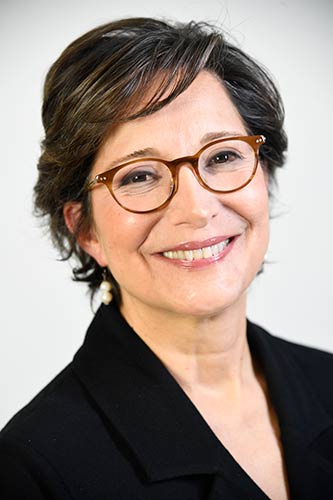
Niloo Ziai, MD
Glaucoma, General Ophthalmology
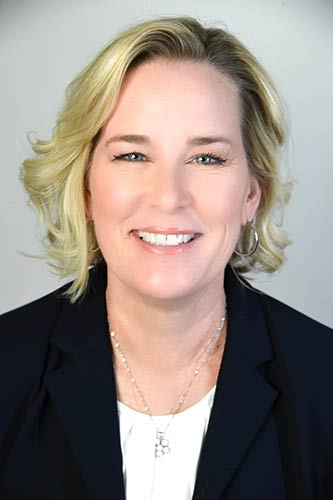
Sarah Merrill, MD
General Opthalmology, Botox
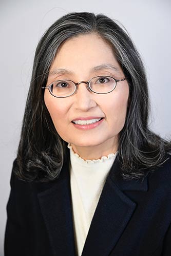
Soo Shin, MD
General Ophthalmology & Corneal Diseases
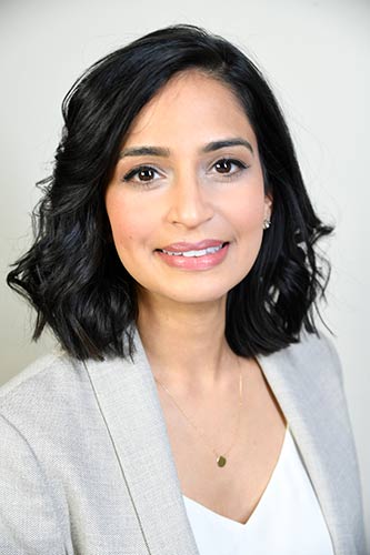
Ruby Parikh, M.D.
General Ophthalmology, Cataract Surgery & Uveitis
Mohib Khan, MD
Glaucoma, Glaucoma Surgery, General Ophthalmology & Cataract Surgery
Christina Chang, MD
General Ophthalmology, Cataract Surgery, Botox
Elaine Bourdeau, OD
Optometry, Contact Lenses
"Techs are very nice, friendly and patient. The doctors are so incredibly smart and explain things so well. They have an answer for every question. Very up to date equipment and technology."

"I found everyone with whom I spoke well informed, helpful and empathetic. Also the brochures provided in the folder I was given, offered very helpful information about the procedure I'm preparing for and eliminated some of my questions. The few remaining questions I asked were thoroughly answered. I like the clarity, good order and expertise I experienced. Something that can't be faked is the enthusiasm and interest of everyone I met. The environment must be a good one in which to work, affirming each person's contribution."
"Everyone was very professional and very kind. I'm so happy to have used their services."
"Your office is very efficient. The staff is extremely friendly and kind, I would definitely recommend your practice to my friends."
Overtime, cataracts can progress to a point where you are no longer able to see clearly and surgery is necessary. Patients will notice blurred vision, trouble seeing at night, light sensitivity and other symptoms which will make driving, reading and enjoyment of recreational activities more challenging. At this time, cataract surgery is reasonable.
Do you think that you might be suffering from cataracts?
Dry eye syndrome is a common disease in which the eye under-produces tears or tears leave the eye too quickly. A normal functioning eye constantly produces tears to form a tear film, which acts as moisturizer and lubricant. For someone with dry eye, the resulting lack of moisture and lubrication can cause a variety of problems.
Do you believe you suffer from Dry Eye?
For patients who wish to look and feel younger, our physicians offer Botox injections. These injections can also help treat certain medical conditions.
Are you interested in learning more about Botox?